
Private Groin & Hernia Ultrasound Scan
Investigating suspected groin hernias, localised swelling and selected abdominal wall hernias.
OUR SERVICE
Are you experiencing a lump, swelling or discomfort in your groin?
A lump or swelling in the groin may be caused by a hernia or another soft tissue abnormality.
Some hernias become more noticeable when standing, coughing, lifting heavy objects or straining, while others may be difficult to identify during a routine physical examination.
At Serenity Ultrasound, we offer private groin and hernia ultrasound scans to investigate suspected hernias and selected causes of localised swelling where ultrasound is clinically appropriate.
What is a hernia?
A hernia occurs when tissue, such as fatty tissue or part of the bowel, pushes through a weakened area of the surrounding muscle or connective tissue.
Hernias can develop in different parts of the body, including the groin and abdominal wall.
Some hernias cause a visible or palpable lump, while others may be associated with discomfort, particularly during physical activity or straining.
What can a groin and hernia ultrasound detect?
Our examination may help assess:
- Inguinal hernia – a hernia occurring in the groin through the inguinal canal.
- Femoral hernia – a hernia occurring in the upper thigh or groin region, below the inguinal ligament.
- Umbilical hernia – a hernia developing around the belly button.
- Paraumbilical hernia – a hernia developing close to the belly button.
- Incisional hernia – a hernia developing through a previous abdominal surgical scar.
- Other abdominal wall hernias – including selected epigastric and Spigelian hernias.
- Muscle hernias – muscle tissue bulging through a weakened area of its surrounding fascia.
- Localised groin swelling – assessment of selected superficial soft tissue abnormalities that may resemble a hernia.
The examination will be tailored to the area of concern and the clinical indication.
When should you consider a groin or hernia ultrasound?
An ultrasound scan may be appropriate if you have:
- An unexplained lump or swelling in your groin.
- A suspected hernia that is not clearly identifiable on physical examination.
- A lump that becomes more noticeable when coughing or straining.
- Localised swelling around your belly button or abdominal wall.
- Swelling near a previous abdominal surgical scar.
- A suspected recurrent hernia following previous surgery.
A hernia that is clearly identifiable during a clinical examination may not require ultrasound confirmation. Your GP or surgeon can advise whether imaging is necessary.
What happens during your scan?
Your sonographer will ask about your symptoms and the location of the suspected hernia.
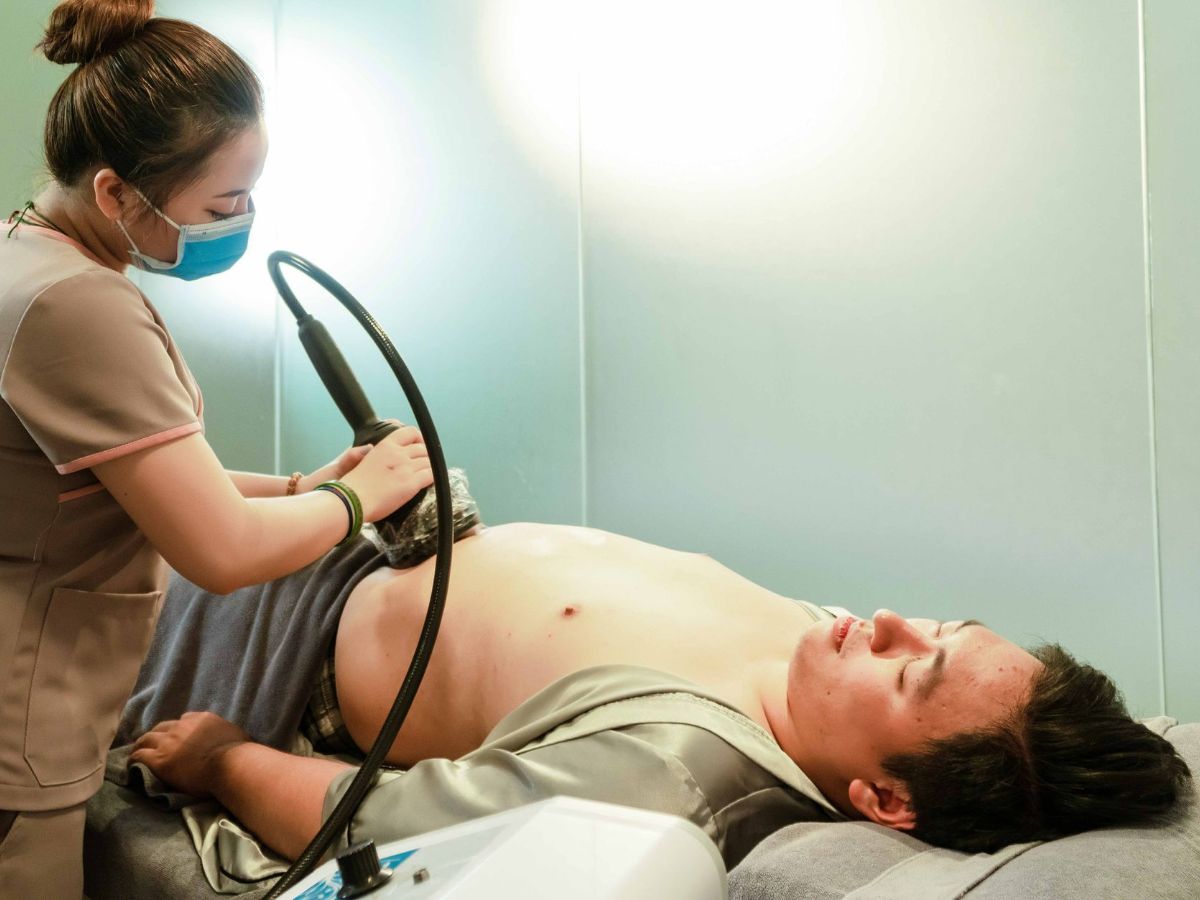
Ultrasound gel will be applied to the affected area, and a handheld probe will be used to examine the underlying tissues.
You may be asked to cough, gently strain or stand during the examination. These movements can help demonstrate a hernia that may not be visible while lying down.
The examination may assess the location of the hernia, the size of the opening and the appearance of any tissue protruding through it.
You will receive a written ultrasound report outlining the findings and any recommended follow-up.
Important: When to seek urgent medical attention
If you have a painful groin or abdominal lump that cannot be pushed back in, particularly if it is associated with severe or worsening pain, vomiting, abdominal swelling or skin discolouration, seek urgent medical assessment.
These symptoms may indicate a trapped or strangulated hernia, which can require emergency treatment.
Do not delay urgent medical care to arrange a routine ultrasound appointment.
Book your private groin and hernia ultrasound scan at Serenity Ultrasound.
OUR PROCESS
Your MSK Ultrasound Appointment at Serenity Ultrasound

1. Your consultation
Your sonographer will ask about your symptoms, medical history and the area of concern to help determine the most appropriate ultrasound assessment.

2. Your ultrasound examination
A small amount of ultrasound gel will be applied to your skin, and a handheld probe will be moved gently over the area being examined. You may be asked to change position or move the affected joint during the scan.

3. Understanding your results
Your sonographer will discuss the examination findings with you and explain whether any abnormalities have been identified.

4. Your report and next steps
You will receive a written ultrasound report detailing the findings. Where appropriate, your sonographer will advise you to seek further assessment or discuss the results with your GP or treating specialist.